Laboratory Tests
Banded karyotype
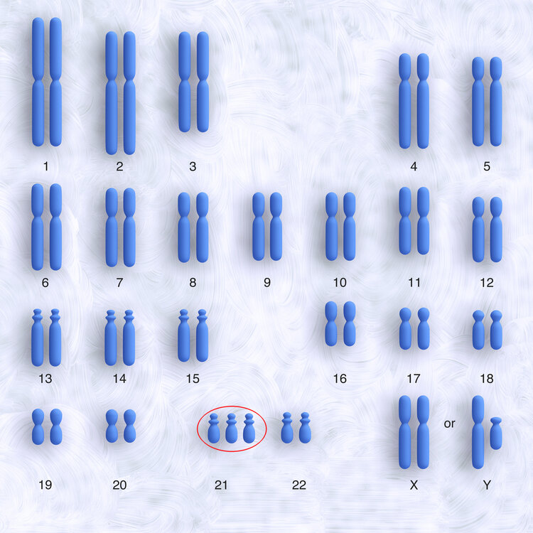
This test has been performed for many years. The cells collected by CVS or amniocentesis are sent to a cytogenetics laboratory. There the cells are cultured (stimulated to grow and divide) for 10-14 days. After enough cells are obtained, a banded karyotype can be performed. This means that the fetal chromosomes in the cultured cells are stained and subsequently photographed. The photographed chromosomes are then ordered by number, counted and checked for structural abnormalities. There should be 46 chromosomes, 23 pairs. A boy's karyotype is described as 46,XY and a girl's karyotype is described as 46,XX. In case of Down Syndrome, there is a third copy of chromosome 21.
It usually takes 2 weeks to get a result of the banded karyotype.
Micro array or molecular karyotype
Microarray, also called molecular karyotype, is a relatively new way of performing detailed testing of the genetic material contained in our chromosomes. Microarray is gradually replacing the banded karyotype.
Every cell in our body contains a full set of our genes on strands of DNA wrapped up in tiny bundles called chromosomes. These thousands of genes are collectively referred to as our genome. Whereas the older chromosome testing technique, banded karyotype, can only detect very large changes in our genome, microarray can detect much smaller variations, usually called 'copy number variants'.
This means that microarray is a very sensitive test which is generally helpful because it is able to detect more causes of developmental and other health problems. The increased sensitivity is occasionally problematic because some 'copy number variants' that are detected are not fully understood or have not been previously reported in the medical literature. In these instances it can be difficult for us to predict what the outcome will be, but testing of the parents and advice from a geneticist will be helpful in some cases.
If it is suspected that your unborn baby might have a chromosomal abnormality we will talk to you and your doctor about testing options, and help you decide whether a microarray or a banded karyotype would be the most appropriate test.
5-probe FISH (Fluorescent in Situ Hybridisation)
A 5-probe FISH can be done in addition to the banded karyotype or micro array.
A 5-probe FISH offers a rapid result, within 24-48 hours for particular chromosomes. It tests for the number of chromosomes 13, 18, 21, X and Y.
Fluorescent stains of different colours are mixed with fresh cells obtained by CVS or amniocentesis. These different coloured stains will bind with chromosome 13, 18, 21, X and Y. The cells are then studied under the fluorescent microscope. In a normal cell there should be 2 fluorescent dots of the colour representing chromosome 13, 2 dots representing chromosome 18, 2 dots representing chromosome 21 and 2 dots representing the X chromosome if the fetus is female and 1 dot for the X chromosome and 1 dot for the Y chromosome if the fetus is male.
In case of trisomy 21 or Down Syndrome, three dots representing chromosome 21 are seen, as in the picture below (the pink dots represent copies of chromosome 21).
There will be an extra charge by the laboratory for a 5-probe FISH.
The 5-probe FISH result will always be followed by a complete banded karyotype or micro array.


